- Zosia Maciorowski
Zosia Maciorowski
Faculty List
- Ali Şengül
- Awtar Ganju-Krishan
- William Telford
- Duygu Sağ
- Elif Çelik
- Elif Karakoç Aydıner
- Esin Çetin Aktaş
- Fatma Betul Oktelik
- Gerhard Wingender
- Günnur Deniz
- Haluk Barbaros Oral
- İhsan Gürsel
- İsmail Cem Yılmaz
- Joanne Lannigan
- Klara Dalva
- Marianna Tzanoudaki
- Mayda Gürsel
- Mirjam van der Burg
- Muzaffer Yıldırım
- Paul Hutchinson
- Paul J. Smith
- Raif Yuecel
- Raquel Cabana
- Sara De Biasi
- Tolga Sütlü
- Uğur Muşabak
- Umut Küçüksezer
- Zeynep Karakan Karakas
- Zosia Maciorowski
Duyurular
Haberler
Webinar
COVID-19
Bilimsel Ödüller
Kongreler
Zosia Maciorowski received her B.Sc. in Microbiology from McGill University in Montreal and M.A. in Biology from Wayne State University in Detroit. She has worked in many labs and countries over the years on a variety of different subjects, from the early days of tissue culture to small animal surgery, monoclonal antibody production and early immunological and molecular biology techniques. In the 80’s she specialized in solid tumor preparation for multicolor and cell cycle analysis. For 28 years she was responsible for the Flow Cytometry Core Facility at the Curie Institute in Paris, France, from which she is now retired. Zosia is Co-Chair of the Live Education Task Force of the International Society for Advancement of Cytometry (ISAC) which has organized international flow cytometry workshops around the world.
Module Descriptions
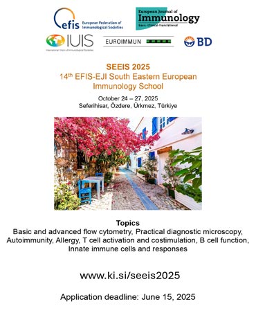
Lecture: Basics of Flow Cytometry This module will familiarize those new to flow cytometry with the basics of fluorescence, flow cytometer hardware components and how a cytometer works. The key components including fluidics, lasers, optics, electronic detectors, analog to digital converters and pulse processors will be described in detail.
Lab: Basic Multicolor Flow Cytometry: Fluorochromes, Spectral Overlap and Compensation
In the lab-based module, participants will learn how calculate stain index in order to rank a variety of fluorochromes based on their brightness and assess spillover problems. Stain Index will be used in the determination of optimal PMT voltage settings and antibody titration. Both automated and manual compensation will be performed, with a discussion of pitfalls and best practices. Compensated single colors will be used to show the effect of spread due to spectral spillover on experimental results. Quality Control and instrument care will also be discussed.
Relevant Literature:
1. Maciorowski, Z., Chattopadhyay, P.K. and Jain, P. 2017. Basic multicolor flow cytometry. Current Protocols in Immunology, 117, 5.4.1-5.4.38.
2. Roederer, M. (2008), How many events is enough? Are you positive?. Cytometry Part A, 73A: 384–385. doi: 10.1002/cyto.a.20549.
3. Leonore A Herzenberg, James Tung, Wayne A Moore, Leonard A Herzenberg & David R Parks. 2008. Interpreting flow cytometry data: a guide for the perplexed. Nature Immunology 7: 7 681-5.

Faculty List
- Ali Şengül
- Awtar Ganju-Krishan
- William Telford
- Duygu Sağ
- Elif Çelik
- Elif Karakoç Aydıner
- Esin Çetin Aktaş
- Fatma Betul Oktelik
- Gerhard Wingender
- Günnur Deniz
- Haluk Barbaros Oral
- İhsan Gürsel
- İsmail Cem Yılmaz
- Joanne Lannigan
- Klara Dalva
- Marianna Tzanoudaki
- Mayda Gürsel
- Mirjam van der Burg
- Muzaffer Yıldırım
- Paul Hutchinson
- Paul J. Smith
- Raif Yuecel
- Raquel Cabana
- Sara De Biasi
- Tolga Sütlü
- Uğur Muşabak
- Umut Küçüksezer
- Zeynep Karakan Karakas
- Zosia Maciorowski
